Contrast–enhanced ultrasound imaging of the optic nerve sheath diameter – a proof of concept study
Prikaz premera ovojnice optičnega živca z ultrazvokom s kontrastnim sredstvom – dokaz zasnove raziskave
DOI:
https://doi.org/10.18690/actabiomed.151Keywords:
eye, magnetic resonance imaging, optic nerve sheath diameter, contrast-enhanced ultrasound, vascular anatomyAbstract
Purpose: The present proof of concept study was performed to evaluate the consistency between measurements of the optic nerve sheath taken using contrast– enhanced sonography and high– resolution magnetic resonance imaging (MRI). The main goal was to devise a novel candidate method to measure the optic nerve sheath diameter that is easy to perform, straightforward to interpret, and highly reproducible.
Methods: The approval of the National Medical Ethics Committee of the Republic of Slovenia was obtained; the study was registered under No. 30/03/12. Nine healthy young adults were examined with high–resolution MRI and contrast–enhanced ultrasound (CEUS). Measurements of the optic nerve sheath diameter were performed 3 mm behind the bulb, using both methods. The datasets were anonymized and the readers, blinded. Statistical analysis included evaluation of agreement using Bland–Altman plots. The assessment of inter– rater reliability was achieved by calculation of the intra class correlation coefficient.
Results: Bland–Altman plots showed favorable overall agreement between both methods, with clinically acceptable limits of agreement. The intra class correlation coefficient calculated for both methods suggests that the CEUS method was at least as reproducible as high–resolution MRI.
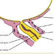
Conclusion: The described method, using CEUS to measure the optic nerve sheath diameter, facilitates the identification of the surrounding anatomy. When CEUS is employed, the measurement points can be easily and reliably set, and retro bulbar artifacts are less confounding. Based on results from a relatively small sample, this method seems to be comparable with high–resolution MRI. Evaluation of the method on a larger sample of healthy subjects and patients with increased intracranial pressure is needed.
Downloads
Downloads
Published
Issue
Section
License
Copyright (c) 2017 Acta Medico-Biotechnica

This work is licensed under a Creative Commons Attribution 4.0 International License.