Radiographic characteristics of impacted teeth
Radiološke značilnosti neizraslih zob
DOI:
https://doi.org/10.18690/actabiomed.113Abstract
Purpose: This study was performed to determine the prevalence, infraosseous position and treatment outcome of impacted teeth in patients treated at our Orthodontic Department over an 11-year period.
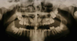
Methods: This retrospective study of orthodontic records was performed on 1,909 patients, who were examined for impacted teeth. It comprised panoramic radiographs, anamnestic and clinical data. We determined the number of subjects with impacted teeth and the number, type and location of the impacted teeth in these subjects. We were also interested in the duration of orthodontic traction with the purpose of bringing the impacted teeth into the dental arch.
Results: Sixty-three (3.3%) out of 1,909 treated orthodontic patients were found to have at least one impacted tooth. A maxillary canine (2.4%) was the most frequently impacted tooth, followed by maxillary and mandibular premolars (0.4%). The majority of patients had one (73%) or two (25.4%) impacted
teeth. Maxillary impacted canines required the longest duration of orthodontic traction.
Conclusion: When orthodontic treatment is performed on patients with impacted teeth, not only the number and the type of the teeth but also the infraosseous position of impacted teeth and their relationship to adjacent structures should be taken into consideration.
Downloads
Downloads
Published
Issue
Section
License
Copyright (c) 2015 Acta Medico-Biotechnica

This work is licensed under a Creative Commons Attribution 4.0 International License.