Follow–up of females with atypical glandular cells on Pap smears
Sledenje žensk z atipičnimi žleznimi celicami v brisih materničnega vratu
DOI:
https://doi.org/10.18690/actabiomed.73Keywords:
cervical cancer screening program, Pap smears, atypical glandular cells, histologic follow– upAbstract
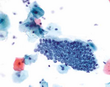
Purpose: Atypical glandular cells (AGC) are often associated with clinically significant lesions, but the interpretation of AGC and the associated lesions on Pap smears remains challenging, even for experienced cytopathologists. The frequency and histologic follow–up of AGC regardless of gland cell type were investigated.
Methods: A retrospective review was conducted involving 199,265 cervicovaginal smears which were examined in the Department of Pathology and Cytology of Celje General Hospital between 1 January 2003 and 31 December 2008. We investigated the frequency of AGC on conventional Pap smears and evaluated the follow–up and correlation between cytology results and subsequent biopsy diagnoses over a 6–year period. The data were processed using the statistics program, SPSS. Statistical significance was set at a p<0.05.
Results: Between 1 January 2003 and 31 December 2008, a total of 745 women had AGC, representing 0.4% of all Pap smear results. One hundred seventy–two women (23.1%) with an average age of 41 years (range, 17–65 years) had a subsequent histologic examination. There were 79 patients (45.9%) with a clinically significant diagnosis, including 63 patients with high– grade cervical intraepithelial neoplasia, 4 patients with adenocarcinoma in situ, and 12 patients with invasive carcinoma (5 endometrial adenocarcinomas, 5 cervical squamous carcinomas, and 2 other types). Normal histologic findings were present in 65 patients (37.8%), 26 patients had low–grade cervical intraepithelial neoplasia, and 2 patients had low–grade glandular intraepithelial neoplasia.
Conclusion: In subsequent biopsy specimens, cytologically AGC are not always glandular or even abnormal. The underlying abnormalities are often so serious that AGC findings require careful clinical and histologic follow–up.
Downloads
Downloads
Published
Issue
Section
License
Copyright (c) 2012 Acta Medico-Biotechnica

This work is licensed under a Creative Commons Attribution 4.0 International License.